Panoramic radiographs are often used in today's dentistry for more accurate diagnosis and treatment planning.
However, 2 dimensional panoramic radiographs are insufficient when 3D investigations are required. 3D dental tomography images taken using a conical beam beam offer great advantages in planning surgical procedures such as implant applications quickly.
Low radiation dose the radiation dose used in Dental CT devices is very low compared to medical CT savers. Radiation SA SA 150 kVp-200ma is applied to image retrieval from the chin and face area with medical CT devices, while in dental CT devices the images are obtained using approximately 80 KVP-5mA.
- Deviations in the measurements made on the panoramic: 3.0 mm-7.5 mm;
- Deviations in periapical measurements: 1.5 mm-5.5 mm and
- Savers in CT measurements: 0.2 mm-0.5 mm
If a measurement with exact values is required, it is recommended to use sails with a material on which a measurement reference can be obtained for CT image retrieval.
Among the cases that can be examined in our patients with this method are:
- Treatment planning before dentomaxillofacial surgery.
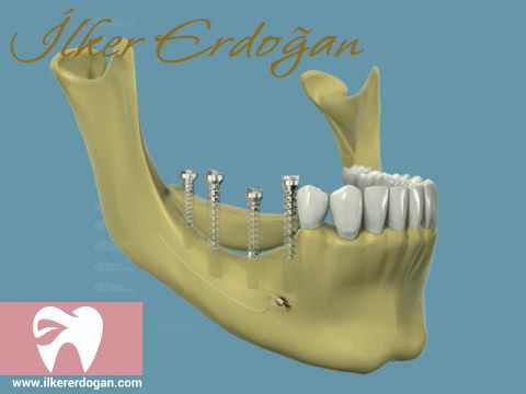
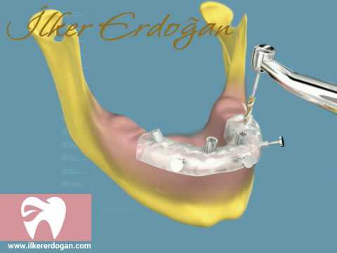
- Preparation of surgical template for Implant placement.
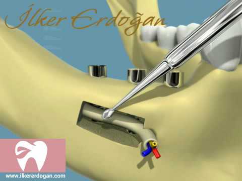
- Examination of anatomical structures such as nasal cavity, incisive canal, maxillary sinus and Mandibular canal.
- Examination of bone quality and density.
- Examination of Jawbone contours.
- Examination of Temporomandibular joint.
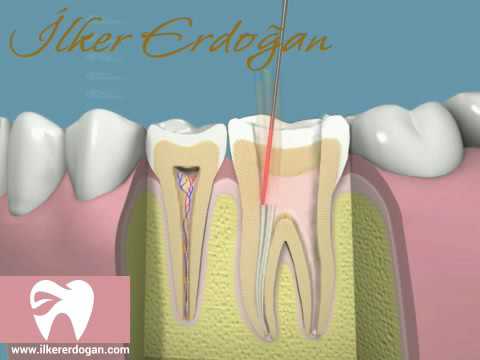
- Examination of periapical area and Canal fillings in root canal treatments.
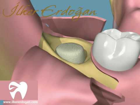
- 3 dimensional analysis of the positions of buried teeth in bone.
- Examination of pathological formations such as cysts and tumours.
- Examination of Root fractures.
- Examination of Periodontal and periapical bone defects.
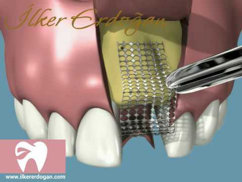
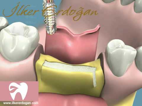
- Planning and follow-up of bone graft applications are involved.


